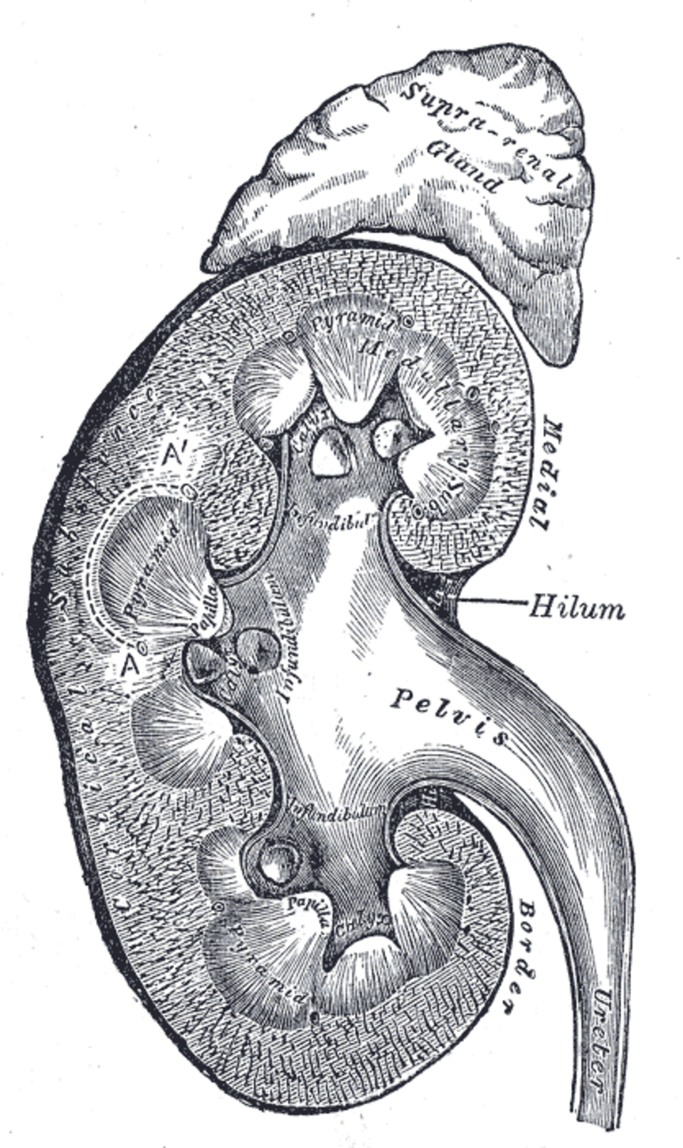
Renal disorders encompass a broad spectrum of conditions affecting the kidneys’ capacity to filter blood, regulate fluid and electrolyte balance, maintain acid-base homeostasis, produce hormones (erythropoietin, renin, calcitriol), and excrete metabolic wastes. Chronic kidney disease affects approximately 10% of the global population and is a leading cause of cardiovascular morbidity and mortality.
Assessment of Kidney Function
Glomerular filtration rate (GFR). The best overall measure of kidney function. Estimated GFR (eGFR) is calculated from serum creatinine using equations (CKD-EPI, MDRD) that account for age, sex, and race. Normal eGFR is >90 mL/min/1.73 m². CKD is defined by eGFR <60 mL/min/1.73 m² or evidence of kidney damage (albuminuria, hematuria, structural abnormalities) for >3 months.
Urinalysis. Dipstick testing detects protein, blood, glucose, leukocytes, and nitrites. Microscopic examination identifies red cells, white cells, casts (hyaline, granular, red cell, white cell), and crystals. Albumin-to-creatinine ratio (ACR) quantifies proteinuria.
Kidney imaging. Renal ultrasound assesses kidney size, cortical thickness, echogenicity, hydronephrosis, cysts, and stones. CT and MRI provide detailed anatomical information. Renal Doppler evaluates renal artery stenosis.
Renal biopsy. Percutaneous needle biopsy is performed for unexplained acute kidney injury, nephrotic syndrome, glomerulonephritis, and transplant dysfunction. Light microscopy, immunofluorescence, and electron microscopy characterize the underlying pathology.
Acute Kidney Injury
AKI is a rapid decline in kidney function over hours to days, manifesting as rise in serum creatinine and/or decrease in urine output. The Kidney Disease: Improving Global Outcomes (KDIGO) classification stages AKI by severity.
Prerenal AKI. Reduced renal perfusion from volume depletion, decreased cardiac output, systemic vasodilation, or renal artery stenosis. The kidney is intrinsically normal. Fractional excretion of sodium (FENa) <1% indicates prerenal physiology. Treatment is volume repletion and optimization of hemodynamics.
Intrinsic AKI. Damage to the renal parenchyma. Acute tubular necrosis (ATN) is the most common cause, resulting from ischemia or nephrotoxins (aminoglycosides, contrast media, myoglobin, cisplatin). Muddy brown granular casts on urinalysis are characteristic. Other causes: acute interstitial nephritis (drug-induced: NSAIDs, antibiotics, PPIs; with eosinophiluria), glomerulonephritis, and vascular causes (renal artery thrombosis, renal vein thrombosis, atheroembolic disease).
Postrenal AKI. Obstruction of the urinary tract (benign prostatic hyperplasia, nephrolithiasis, retroperitoneal fibrosis, malignancy). Anuria alternating with polyuria is suggestive. Renal ultrasound shows hydronephrosis. Relief of obstruction (catheter, nephrostomy, stent) reverses AKI if intervention is timely.
Chronic Kidney Disease
CKD is defined by structural or functional kidney abnormalities persisting for >3 months. Stages 1–5 are based on eGFR. The leading causes are diabetes mellitus (40% of incident ESRD) and hypertension (25%). Other causes include glomerulonephritis, polycystic kidney disease, and interstitial nephritis.
Pathophysiology. Hyperfiltration injury — the remaining nephrons undergo compensatory hyperfiltration and hypertrophy, which over time causes glomerulosclerosis and progressive kidney function decline. Proteinuria is both a marker of injury and a mediator of further damage.
Management. Slowing progression: blood pressure control (ACE inhibitors or ARBs as first-line for proteinuric CKD), glycemic control, dietary protein restriction, and avoidance of nephrotoxins. Managing complications: anemia (erythropoiesis-stimulating agents, iron supplementation), mineral and bone disorder (phosphate binders, vitamin D analogs, calcimimetics), metabolic acidosis (sodium bicarbonate), and cardiovascular risk reduction (statins, antiplatelets).
Glomerular Diseases
Nephrotic Syndrome
Nephrotic syndrome is defined by massive proteinuria (>3.5 g/day), hypoalbuminemia, edema, and hyperlipidemia.
Minimal change disease (MCD). The most common cause of nephrotic syndrome in children. Light microscopy is normal; electron microscopy shows diffuse foot process effacement. Highly responsive to corticosteroids.
Focal segmental glomerulosclerosis (FSGS). A podocyte disease causing sclerosis of some glomerular segments. Primary FSGS responds variably to immunosuppression. Secondary FSGS results from hyperfiltration (obesity, reflux nephropathy, sickle cell disease).
Membranous nephropathy. The most common cause of nephrotic syndrome in adults (non-diabetic). Autoantibodies to phospholipase A2 receptor (PLA2R) are present in 70% of cases. Spontaneous remission occurs in one-third. Immunosuppression with rituximab, cyclophosphamide, or calcineurin inhibitors is used for progressive disease.
Nephritic Syndrome
Nephritic syndrome presents with hematuria (often with red cell casts), hypertension, mild proteinuria, and oliguria.
Post-infectious glomerulonephritis. Immune complex deposition after infection (typically Group A streptococcal pharyngitis or impetigo). Presents 1–3 weeks after infection. Most cases resolve spontaneously.
IgA nephropathy (Berger disease). The most common glomerulonephritis worldwide. Mesangial IgA deposition causes recurrent hematuria (often coincident with mucosal infections). Progressive disease is treated with ACE inhibitors, corticosteroids, and immunosuppression. IgA vasculitis (Henoch-Schönlein purpura) is the systemic form.
Lupus nephritis. A major complication of systemic lupus erythematosus. The ISN/RPS classification (Classes I–VI) guides treatment. Class III and IV (proliferative) lupus nephritis requires aggressive immunosuppression with mycophenolate mofetil or cyclophosphamide.
Rapidly Progressive Glomerulonephritis
RPGN is characterized by crescent formation on biopsy and rapid loss of kidney function over weeks to months. Causes include anti-glomerular basement membrane disease (Goodpasture syndrome), ANCA-associated vasculitis (microscopic polyangiitis, granulomatosis with polyangiitis), and immune complex disease. Treatment is high-dose corticosteroids, cyclophosphamide or rituximab, and plasma exchange (for anti-GBM disease).
Kidney Stones (Nephrolithiasis)
Prevalence is increasing globally. Calcium oxalate stones are most common (60–70%). Other types: calcium phosphate, uric acid, struvite (infection-related), and cystine. Risk factors include low urine volume, hypercalciuria, hyperoxaluria, hyperuricosuria, hypocitraturia, and urinary tract infections.
Clinical presentation. Sudden, severe flank pain radiating to the groin (renal colic), hematuria, nausea, and vomiting. CT without contrast is the diagnostic gold standard.
Management. Acute: hydration, NSAIDs, alpha-blockers (tamsulosin) for medical expulsive therapy of distal ureteral stones, and urgent urologic intervention for obstructed, infected, or large stones. Preventive: increased fluid intake, dietary modifications, and pharmacotherapy based on stone composition and metabolic workup.
Polycystic Kidney Disease
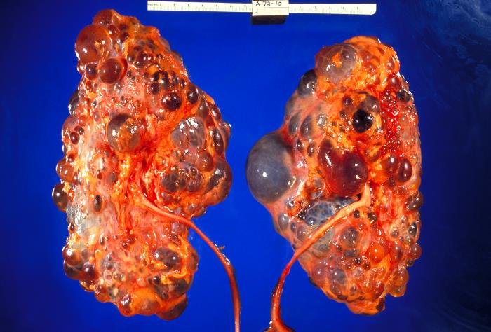
Autosomal dominant PKD (ADPKD) is the most common inherited kidney disease, caused by mutations in PKD1 or PKD2. Multiple cysts progressively enlarge the kidneys, causing pain, hypertension, hematuria, infection, and eventual ESRD (typically in the 5th–6th decade). Tolvaptan (a vasopressin V2 receptor antagonist) slows cyst growth and kidney function decline. Endovascular embolization or nephrectomy for massive kidneys.
End-Stage Renal Disease and Renal Replacement Therapy
ESRD (CKD stage 5, eGFR <15) requires renal replacement therapy for survival.
Hemodialysis. Blood is circulated through a dialyzer (artificial kidney) to remove wastes and excess fluid, typically 3 times per week for 3–4 hours. Vascular access via arteriovenous fistula (preferred), AV graft, or central venous catheter.
Peritoneal dialysis. The peritoneal membrane serves as the filter. Dialysate is instilled into the peritoneal cavity via a catheter, dwells for 4–6 hours, and drains. Advantages include flexibility and better preservation of residual kidney function.
Kidney transplantation. The optimal therapy for ESRD, offering improved survival and quality of life compared to dialysis. Living donor transplantation has superior outcomes. Immunosuppression (calcineurin inhibitors, mycophenolate, corticosteroids) prevents rejection. Transplant recipients face risks of acute and chronic rejection, infection, and cardiovascular disease.
Summary
Renal disorders span acute (AKI) to chronic (CKD) presentations, with etiologies ranging from prerenal and postrenal causes to intrinsic glomerular, tubular, and vascular diseases. CKD is a global epidemic driven by diabetes and hypertension. Glomerulonephritis requires precise histological diagnosis for appropriate immunosuppression. Kidney transplantation offers the best outcomes for ESRD. Across all renal disorders, blood pressure control, proteinuria reduction, and avoidance of nephrotoxins are foundational to preserving kidney function.