The urinary system filters the blood to remove waste products, regulates fluid and electrolyte balance, maintains acid-base homeostasis, and eliminates waste as urine. The kidneys perform the filtration and regulation; the ureters, bladder, and urethra store and transport the urine.
Kidneys
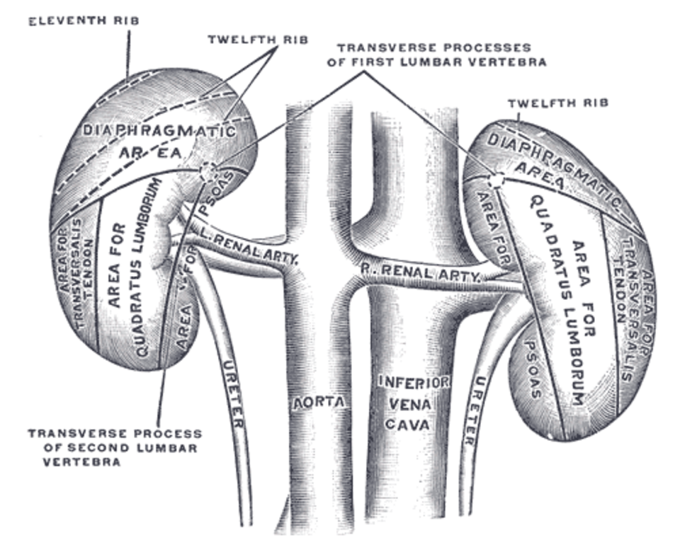
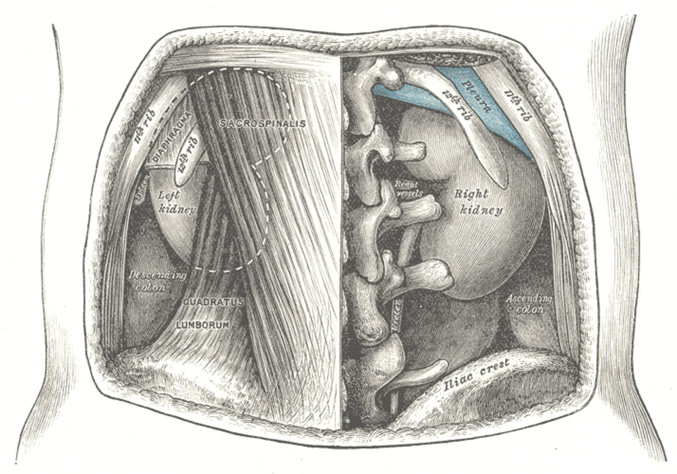
The kidneys are paired, bean-shaped organs approximately 11 cm long, 6 cm wide, and 3 cm thick. They lie on the posterior abdominal wall, retroperitoneally, at the level of T12–L3. The right kidney is slightly lower than the left due to the liver.
External Anatomy
Each kidney is covered by three layers:
- Renal capsule. A tough fibrous layer that directly covers the kidney and maintains its shape.
- Perirenal fat (adipose capsule). A layer of adipose tissue that cushions the kidney.
- Renal fascia. Fibrous connective tissue that anchors the kidney to surrounding structures.
The medial border has a concave notch called the renal hilum, through which the renal artery, renal vein, renal pelvis, lymphatics, and nerves enter and exit.
Internal Anatomy
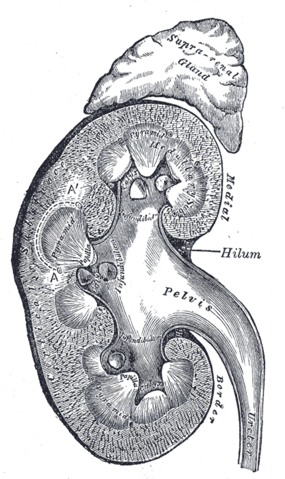
A frontal section through the kidney reveals two distinct regions:
Renal cortex. The outer region (approximately 1 cm thick), containing the renal corpuscles (glomeruli) and convoluted tubules. Cortical columns (columns of Bertin) extend between the renal pyramids.
Renal medulla. The inner region, consisting of 8–18 cone-shaped renal pyramids. The base of each pyramid faces the cortex; the apex (renal papilla) projects into the minor calyx. The medulla contains the loops of Henle and collecting ducts.
Calyces and renal pelvis. Urine drains from the renal papilla into a minor calyx (a cup-shaped collecting structure). Several minor calyces unite to form a major calyx. Two or three major calyces unite to form the renal pelvis, a funnel-shaped structure that exits at the hilum and becomes the ureter.
Blood Supply of the Kidney
The kidneys receive approximately 20–25% of cardiac output (1,200 mL/min) despite constituting only 0.5% of body mass. The blood supply is essential for both oxygen delivery and glomerular filtration.
The renal artery branches sequentially:
- Segmental arteries (5 branches)
- Interlobar arteries (between pyramids)
- Arcuate arteries (at the corticomedullary junction)
- Interlobular (cortical radiate) arteries
- Afferent arterioles → glomerular capillaries → efferent arterioles
- Peritubular capillaries (surrounding the tubules) or vasa recta (descending into the medulla)
The vasa recta are long, straight capillary loops that descend alongside the loops of Henle and collecting ducts. They play a critical role in maintaining the medullary osmotic gradient required for urine concentration.
The Nephron
The nephron is the functional unit of the kidney. Each kidney contains approximately 1–1.5 million nephrons. Each nephron consists of a renal corpuscle (where filtration occurs) and a renal tubule (where reabsorption and secretion occur).
Renal Corpuscle
The renal corpuscle consists of the glomerulus (a tuft of capillaries) surrounded by Bowman capsule (a double-walled epithelial cup). Blood enters the glomerulus through the afferent arteriole and leaves through the efferent arteriole.
The filtration barrier has three layers:
- Fenestrated glomerular capillary endothelium. Pores (70–100 nm) exclude blood cells but allow plasma proteins.
- Glomerular basement membrane. A negatively charged meshwork of collagen and proteoglycans that excludes larger proteins (albumin, > 60 kDa).
- Podocyte foot processes (visceral layer of Bowman capsule). Interdigitating foot processes (pedicels) with filtration slits (20–30 nm) bridged by slit diaphragms (nephrin).
The filtration barrier is highly selective: water, small solutes (glucose, electrolytes, urea), and small proteins pass freely, while blood cells and most plasma proteins are retained. The glomerular filtration rate (GFR) averages 125 mL/min (180 L/day), of which 99% is reabsorbed.
Juxtaglomerular Apparatus
At the vascular pole of each glomerulus, the afferent arteriole contains specialized smooth muscle cells called juxtaglomerular (JG) cells that secrete renin in response to low blood pressure, low sodium delivery, or sympathetic stimulation. A region of the distal tubule (the macula densa) senses sodium chloride concentration and signals the JG cells. This juxtaglomerular apparatus regulates GFR and blood pressure through the renin-angiotensin-aldosterone system (RAAS).
Renal Tubule
The filtrate from Bowman space enters the proximal convoluted tubule and then passes through:
Proximal convoluted tubule (PCT). The longest and most metabolically active segment. It reabsorbs approximately 65% of filtered water, sodium, and chloride, and nearly 100% of filtered glucose and amino acids. It also secretes hydrogen ions (contributing to acid-base balance) and organic cations/anions (drugs, toxins).
Loop of Henle. A U-shaped tubule with descending and ascending limbs. It establishes the medullary osmotic gradient essential for water concentration:
- Thin descending limb. Permeable to water, impermeable to solutes. Water leaves by osmosis, concentrating the tubular fluid.
- Thin ascending limb. Impermeable to water, permeable to solutes. Sodium and chloride diffuse out.
- Thick ascending limb. Impermeable to water; actively reabsorbs sodium, potassium, and chloride via the Na-K-2Cl cotransporter (NKCC2). This is the target of loop diuretics (furosemide).
Distal convoluted tubule (DCT). Reabsorbs sodium via the Na-Cl cotransporter (target of thiazide diuretics) and calcium under the influence of parathyroid hormone. It is also involved in potassium secretion.
Collecting duct. Passes through the medulla to the renal papilla. Its permeability to water is controlled by antidiuretic hormone (ADH). With ADH, aquaporins are inserted into the apical membrane, allowing water reabsorption and producing concentrated urine. Without ADH, the duct is impermeable to water, producing dilute urine. The collecting duct is also the site of aldosterone action (sodium reabsorption and potassium secretion) and acid secretion (intercalated cells).
Ureters
Each ureter is a muscular tube (25–30 cm long) that conveys urine from the renal pelvis to the bladder by peristaltic contractions (every 10–30 seconds). The ureter descends retroperitoneally, crossing the pelvic brim at the bifurcation of the common iliac artery.
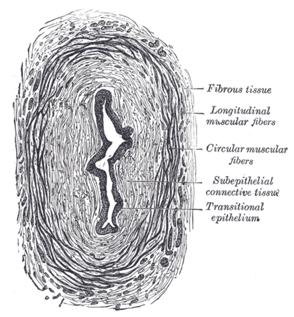
The ureter has three layers:
- Mucosa. Lined by transitional epithelium (urothelium), which can stretch and accommodate varying volumes. It has a star-shaped lumen in cross-section.
- Muscularis. Smooth muscle arranged in inner longitudinal and outer circular layers (a third longitudinal layer appears in the lower third).
- Adventitia. Fibrous connective tissue with blood vessels, lymphatics, and nerves.
The ureter enters the bladder wall obliquely, creating a valve-like mechanism that prevents vesicoureteral reflux (backflow of urine from the bladder into the ureter).
Urinary Bladder
The bladder is a hollow, distensible muscular organ that stores urine. When empty, it lies in the pelvis and has a pyramidal shape. As it fills, it rises into the abdomen. Maximum capacity is approximately 400–600 mL, though the urge to void occurs at about 200–300 mL.
The bladder has four regions:
- Apex (vertex). The anterior tip, attached to the median umbilical ligament (urachal remnant).
- Fundus (base). The posterior wall, containing the two ureteric orifices and the internal urethral orifice.
- Body. The main part between the apex and fundus.
- Neck. The narrowing funnel at the base, leading to the urethra.
Trigone. A smooth, triangular region on the internal floor of the bladder, bounded by the two ureteric orifices and the internal urethral orifice. The trigone is clinically significant because it is the last area to stretch (so it is the most sensitive to distension) and the most common site for infection and tumors.
Bladder wall layers:
- Mucosa. Transitional epithelium (urothelium). In an empty bladder, the epithelium is thick (5–6 cell layers) and folded into rugae. As the bladder fills, the epithelium stretches to 2–3 layers.
- Lamina propria. Connective tissue layer beneath the mucosa.
- Muscularis (detrusor muscle). Three layers of smooth muscle (inner and outer longitudinal, middle circular). The detrusor contracts during urination.
- Serosa (peritoneum). Covers the superior surface; adventitia on other surfaces.
Urethra
Male urethra (18–20 cm). Serves both urinary and reproductive functions. It has three segments:
- Prostatic urethra (3–4 cm). Passes through the prostate; receives the ejaculatory ducts and the prostatic ducts.
- Membranous urethra (1–2 cm). Passes through the urogenital diaphragm; the narrowest and least distensible segment.
- Spongy (penile) urethra (15 cm). Passes through the corpus spongiosum; ends at the external urethral orifice.
Female urethra (3–4 cm). Shorter than the male urethra, running from the bladder neck to the external urethral orifice in the vestibule. Its shortness makes females more susceptible to urinary tract infections.
Sphincters
- Internal urethral sphincter. At the bladder neck; composed of smooth muscle (detrusor thickening). Under involuntary (sympathetic) control; maintains urinary continence.
- External urethral sphincter. In the urogenital diaphragm; composed of skeletal muscle (rhabdosphincter). Under voluntary (somatic) control via the pudendal nerve. It allows voluntary interruption of urination.
Micturition (Urination)
Micturition is the process of emptying the bladder. It is controlled by a complex reflex coordinated in the pontine micturition center (Barrington nucleus) and modulated by voluntary cortical input.
Filling phase. The detrusor muscle is relaxed (sympathetic-mediated, β₃-adrenergic) and the internal sphincter is contracted (sympathetic-mediated, α₁-adrenergic). As the bladder fills, stretch receptors in the detrusor send afferent signals to the spinal cord (pelvic nerves, S2–S4).
Voiding phase. When bladder volume reaches approximately 200–300 mL, the pontine micturition center is activated. It sends parasympathetic signals (pelvic nerves, acetylcholine, M₃ receptors) to contract the detrusor and relax the internal sphincter. Simultaneously, somatic signals to the external sphincter are inhibited, allowing relaxation and urine flow.
Cortical input can suppress the micturition reflex voluntarily (maintaining continence despite a full bladder) or facilitate it (voiding before the bladder is full). This voluntary control develops at approximately 2–3 years of age.
Summary
The urinary system filters the blood through approximately 2 million nephrons, reabsorbing essential substances and excreting waste. The nephron’s sophisticated countercurrent system allows the kidney to produce urine ranging from dilute to highly concentrated as needed. The ureters transport urine by peristalsis, the bladder stores it, and coordinated neural reflexes control micturition. Understanding the anatomy of the urinary tract is essential for diagnosing and treating common conditions including infection, stones, obstruction, and kidney disease.