Overview
The skeletal system is the body’s structural framework — a living, dynamic organ system that does far more than hold us upright. It protects the soft organs inside us, works with muscles to produce movement, stores essential minerals, and houses the marrow that manufactures our blood cells. Far from being a collection of dead, dry sticks, our bones are richly supplied with blood vessels and nerves, constantly being remodeled and repaired.
If you think of the skeleton as the body’s scaffolding, you have the right idea — but scaffolding is static and passive. The skeleton is neither. It grows with you through childhood, strengthens with exercise, repairs itself when broken, and gradually loses density with age. Every bone in your body today is not the same bone you had a decade ago; it has been partially taken apart and rebuilt through the continuous process of remodeling.
Functions of the Skeleton
The skeleton performs six essential functions that span mechanical support, protection, movement, mineral storage, blood cell production, and energy storage:
Support — Bones form a rigid framework that holds the body upright and provides attachment points for soft tissues. Without the skeleton, the body would collapse under its own weight.
Protection — The skeleton encloses and shields the body’s most vulnerable organs. The skull cradles the brain, the vertebral column surrounds the spinal cord, and the rib cage guards the heart, lungs, and major vessels.
Movement — Bones act as levers that muscles pull against to produce motion. Joints between bones allow different types and ranges of movement, from the subtle gliding of wrist bones to the powerful rotation of the hip.
Mineral storage — Bone tissue serves as the body’s primary mineral reservoir, holding approximately 99% of total body calcium and 85% of phosphorus. These minerals can be released into the bloodstream as needed to maintain critical physiological concentrations.
Hematopoiesis — Red bone marrow, located primarily in the axial skeleton of adults, produces all types of blood cells: red blood cells (erythrocytes), white blood cells (leukocytes), and platelets (thrombocytes).
Energy storage — Yellow marrow, which replaces red marrow in the shafts of long bones with age, consists primarily of adipose tissue and serves as an energy reserve.
| Function | Key Role |
|---|---|
| Support | Structural framework, soft tissue attachment |
| Protection | Encloses brain, spinal cord, thoracic organs |
| Movement | Lever system for muscle action |
| Mineral storage | Calcium (99%), phosphorus (85%) reservoir |
| Hematopoiesis | Blood cell production in red marrow |
| Energy storage | Yellow marrow (adipose tissue) |
Classification of Bones by Shape
Bones are classified into six categories based on their shape. This classification reflects not only their appearance but also their mechanical function and developmental origin.
Long bones are longer than they are wide, consisting of a shaft (diaphysis) with two expanded ends (epiphyses). They function as levers and are found in the limbs: femur, tibia, fibula, humerus, radius, ulna, and the phalanges. Despite their name, long bones are defined by shape, not absolute size — the small bones of the fingers qualify as long bones.
Short bones are approximately cube-shaped, with length and width roughly equal. They provide stability with limited motion and are found in the wrist (carpals) and ankle (tarsals). The patella, though often classified with sesamoid bones, also has short bone characteristics.
Flat bones are thin, flattened, and usually curved. They provide extensive surfaces for muscle attachment and enclose vital organs. Examples include the cranial bones (frontal, parietal, occipital), the sternum, ribs, and scapulae.
Irregular bones have complex shapes that do not fit into the other categories. Their elaborate structure reflects specialized functions such as protecting the spinal cord (vertebrae) or supporting the jaw (mandible). The sacrum, coccyx, and pelvic bones also fall into this group.
Sesamoid bones develop within tendons where they cross joints, protecting the tendon from compressive forces and improving mechanical leverage. The patella is the largest sesamoid bone. The pisiform of the wrist is another example. Sesamoid number varies between individuals.
Sutural (Wormian) bones are small, irregular bones that occasionally form within the sutures of the skull. Their presence and number are variable and of no clinical significance in most cases, though they may indicate underlying developmental conditions when numerous.
Macroscopic Bone Structure
A typical long bone — such as the humerus or femur — reveals the fundamental organizational plan of bone at the gross anatomical level.
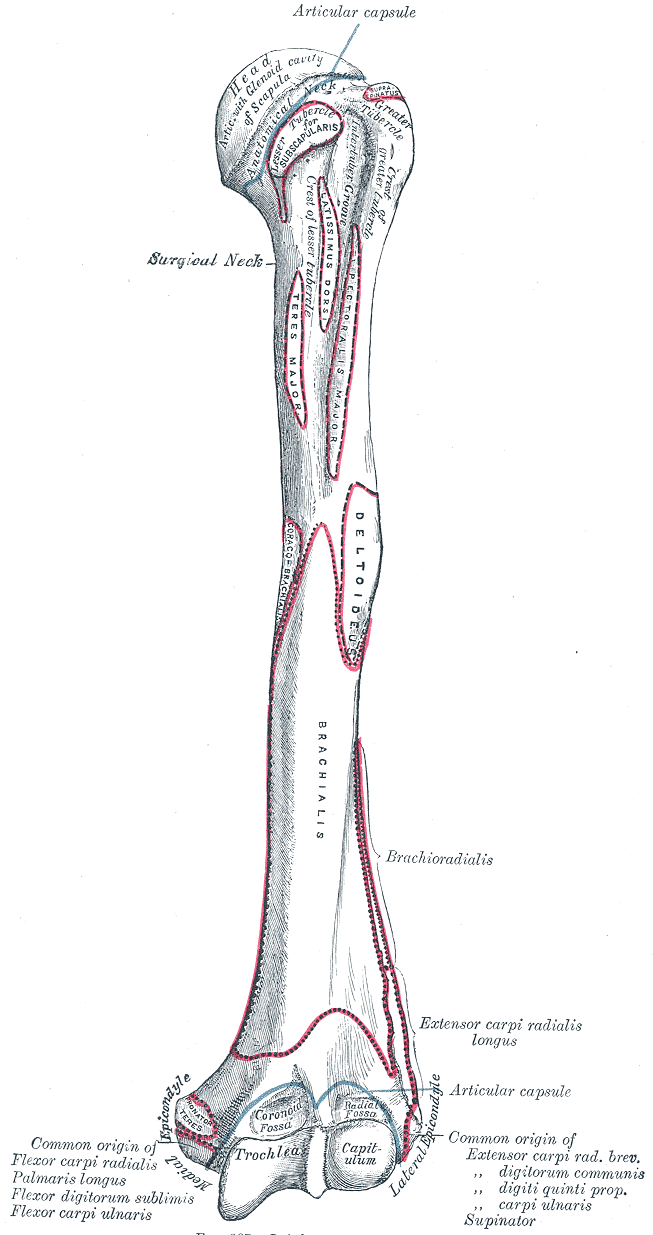
The diaphysis is the elongated shaft of the bone, composed primarily of compact bone surrounding a central medullary cavity. The thick cortical wall of the diaphysis provides the bone’s main load-bearing capacity. The medullary cavity within contains bone marrow and is lined by a thin membrane called the endosteum.
The epiphyses are the expanded ends of the bone, found proximal and distal to the diaphysis. They consist of a thin outer shell of compact bone surrounding an interior of cancellous (trabecular) bone. The joint surfaces of the epiphyses are covered by articular cartilage, a smooth layer of hyaline cartilage that reduces friction during movement.
The metaphysis is the region between the diaphysis and each epiphysis. In growing bones, this area contains the epiphyseal (growth) plate — a cartilaginous disc where longitudinal bone growth occurs. After growth ceases, the epiphyseal plate is replaced by a bony remnant called the epiphyseal line.
The periosteum is a dense, fibrous connective tissue membrane that covers the external surface of bone except where the articular cartilage is present. It has two layers: an outer fibrous layer of dense irregular connective tissue and an inner osteogenic layer containing osteoprogenitor cells. The periosteum is richly supplied with blood vessels and nerves, which is why bone injuries are so painful and why periosteal blood supply is critical for fracture healing. Sharpey’s fibers (collagen bundles) anchor the periosteum firmly to the underlying bone.
The endosteum is a thin membrane lining the internal surfaces of bone — the medullary cavity and the trabeculae of cancellous bone. Like the periosteum’s inner layer, the endosteum contains osteoprogenitor cells, osteoblasts, and osteoclasts, making it an active participant in bone growth, remodeling, and repair.
| Region | Composition | Key Feature |
|---|---|---|
| Diaphysis | Compact bone (thick cortical layer) | Shaft; medullary cavity |
| Epiphysis | Cancellous bone with compact shell | Expanded end; articular cartilage |
| Metaphysis | Transitional zone | Growth plate (children) |
| Periosteum | Fibrous + osteogenic layers | External covering; pain-sensitive |
| Endosteum | Single cell layer | Internal lining; remodeling |
Microscopic Bone Structure
At the microscopic level, bone tissue exists in two forms: compact (cortical) bone and cancellous (trabecular) bone. They share the same matrix composition and cell types but differ dramatically in their architectural organization.
Compact Bone and the Osteon
Compact bone forms the dense outer wall of all bones and constitutes approximately 80% of total bone mass. It is organized into repeating structural units called osteons (Haversian systems).
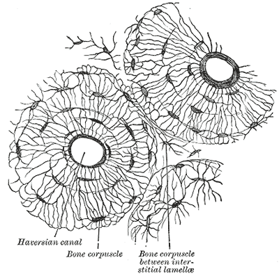
Each osteon is a cylindrical structure aligned parallel to the bone’s long axis. At its center is the Haversian canal, which contains blood vessels, nerves, and lymphatic vessels. Surrounding the central canal are concentric rings of mineralized matrix called concentric lamellae, like the layers of an onion. Between the lamellae are small spaces called lacunae, each housing an osteocyte (a mature bone cell). Radiating from each lacuna are tiny channels called canaliculi, which connect osteocytes to each other and to the blood supply, allowing the exchange of nutrients and waste.
Between osteons lie interstitial lamellae — remnants of older osteons that were partially destroyed during bone remodeling. Along the inner and outer surfaces of the bone are circumferential lamellae, which run the entire circumference of the bone and provide additional strength.
The organization of compact bone into osteons gives it remarkable strength for its weight. The concentric lamellae resist compressive forces, while the collagen fibers within each lamella are oriented in alternating directions, resisting torsional and tensile forces from multiple angles.
| Structure | Description |
|---|---|
| Haversian canal | Central channel; contains vessels and nerves |
| Concentric lamellae | Rings of mineralized matrix around central canal |
| Lacunae | Spaces between lamellae; house osteocytes |
| Canaliculi | Microscopic channels connecting lacunae |
| Interstitial lamellae | Remnants of old, partially resorbed osteons |
| Circumferential lamellae | Outer and inner layers of compact bone |
Cancellous (Trabecular) Bone
Cancellous bone, also called trabecular or spongy bone, forms the interior of most bones and constitutes approximately 20% of total bone mass. Unlike the dense, organized structure of compact bone, cancellous bone consists of a lattice-like network of thin, branching plates and rods called trabeculae.
Trabeculae are not arranged randomly. They align precisely along the lines of mechanical stress that a bone experiences during normal loading — a phenomenon described by Wolff’s law, which states that bone adapts to the loads placed upon it. This trabecular orientation provides maximum strength with minimum weight.
The spaces between trabeculae contain bone marrow (red or yellow) and blood vessels. Cancellous bone does not contain true osteons. Instead, the trabeculae are covered by a thin layer of endosteum containing osteoblasts and osteoclasts. Because of its large surface area relative to its volume, cancellous bone has a much higher metabolic turnover rate than compact bone — approximately five times faster. This makes it more responsive to hormonal stimuli but also more vulnerable to the bone loss of osteoporosis.
Bone Cells
Bone is populated by four types of cells, each derived from a distinct lineage and serving a specialized function. Together, they maintain the bone as a living, dynamic tissue.
Osteoprogenitor cells are mesenchymal stem cells that give rise to osteoblasts. They are found in the periosteum, endosteum, and bone marrow. When stimulated — by growth factors, mechanical stress, or fracture — they proliferate and differentiate into osteoblasts.
Osteoblasts are the bone-forming cells. They synthesize and secrete the organic components of bone matrix (osteoid), including type I collagen and proteoglycans. Osteoblasts are plump, cuboidal cells arranged in a single layer on bone surfaces. They deposit osteoid and then regulate its subsequent mineralization by releasing matrix vesicles that concentrate calcium and phosphate. Once surrounded by matrix, osteoblasts differentiate further into osteocytes or undergo apoptosis.
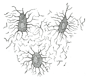
Osteocytes are mature bone cells that originate from osteoblasts trapped within the matrix they secreted. They are the most abundant bone cell type. Each osteocyte resides in a lacuna and extends long cytoplasmic processes through canaliculi to contact neighboring osteocytes. This interconnected network senses mechanical load and orchestrates the bone’s adaptive response. Osteocytes can signal osteoblasts to form bone where it is needed and osteoclasts to resorb bone where it is not.
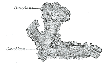
Osteoclasts are large, multinucleated cells responsible for bone resorption. Unlike the other bone cells, which derive from mesenchymal stem cells, osteoclasts originate from the fusion of hematopoietic stem cells of the monocyte-macrophage lineage. They attach to the bone surface and create a sealed-off compartment (How ship lacuna) into which they secrete hydrogen ions and proteolytic enzymes that dissolve the mineral and digest the organic matrix. Osteoclast activity is regulated by hormones such as parathyroid hormone (stimulates) and calcitonin (inhibits).
| Cell | Origin | Function | Location |
|---|---|---|---|
| Osteoprogenitor | Mesenchymal stem cell | Differentiate into osteoblasts | Periosteum, endosteum, marrow |
| Osteoblast | Osteoprogenitor | Bone formation (osteoid deposition, mineralization) | Bone surface (active) |
| Osteocyte | Osteoblast (mature) | Mechanosensing, matrix maintenance, signaling | Lacunae |
| Osteoclast | Hematopoietic (monocyte lineage) | Bone resorption (breakdown) | How ship lacunae on bone surface |
Bone Matrix
Bone matrix is a composite material whose properties arise from the combination of inorganic (mineral) and organic components. Like reinforced concrete, the mineral provides compressive strength while the organic component provides tensile strength and flexibility.
The inorganic component accounts for approximately 65% of bone’s dry weight. It consists primarily of hydroxyapatite — crystalline calcium phosphate (Ca₁₀(PO₄)₆(OH)₂) — along with smaller amounts of carbonate, magnesium, fluoride, and other ions. These mineral crystals are deposited in and around the collagen fibrils of the organic matrix, giving bone its hardness and resistance to compression.
The organic component (osteoid) makes up approximately 35% of bone’s dry weight. Type I collagen accounts for 90% of the organic matrix, providing tensile strength and the scaffold upon which mineralization occurs. The remaining 10% includes proteoglycans, glycoproteins (osteonectin, osteocalcin, osteopontin), and growth factors. These non-collagenous proteins regulate matrix mineralization, cell attachment, and signaling.
| Component | Percentage | Function |
|---|---|---|
| Hydroxyapatite | 65% | Compressive strength, hardness |
| Collagen type I | ~31% | Tensile strength, flexibility |
| Other proteins | ~4% | Mineralization regulation, cell signaling |
Bone Development and Growth
Intramembranous Ossification
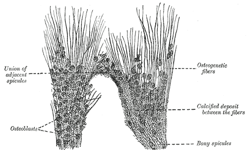
Intramembranous ossification is the process by which bone forms directly within mesenchymal connective tissue, without a preceding cartilage model. This process produces the flat bones of the skull (frontal, parietal, parts of the occipital and temporal), portions of the mandible and clavicle, and contributes to bone thickening throughout life.
The process begins when mesenchymal cells condense and differentiate into osteoblasts at ossification centers. These osteoblasts secrete osteoid, which quickly mineralizes, trapping some osteoblasts as osteocytes. The resulting bone spicules radiate outward from the ossification centers, forming trabeculae that become cancellous bone. The surrounding mesenchyme differentiates into the periosteum, and the outer layers of the developing bone are remodeled into compact bone.
Endochondral Ossification
Endochondral ossification is the process by which bone replaces a cartilage model. This is how most bones of the skeleton form, including the long bones of the limbs, vertebrae, ribs, and the base of the skull.
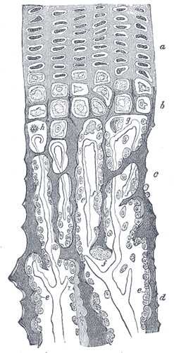
The process begins in the fetus when mesenchymal cells condense and differentiate into chondrocytes, forming a hyaline cartilage model shaped like the future bone. A periosteal bone collar forms around the diaphysis of the cartilage model, and the cartilage within becomes calcified. Blood vessels invade, carrying osteoblasts and osteoclasts. The osteoblasts deposit bone on the calcified cartilage remnants, forming a primary ossification center in the diaphysis. After birth, secondary ossification centers appear in the epiphyses.
Growth Plate
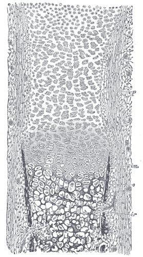
In growing bones, the epiphyseal (growth) plate separates the diaphysis from each epiphysis. This cartilaginous disc is the site of longitudinal bone growth. The growth plate is organized into five zones that reflect the progressive maturation and replacement of cartilage by bone:
- Resting (reserve) zone — Small, inactive chondrocytes anchor the plate to the epiphysis.
- Proliferative zone — Chondrocytes divide and stack in columns, producing new cartilage matrix.
- Hypertrophic zone — Chondrocytes enlarge and the matrix between them becomes calcified.
- Calcification zone — Matrix mineralization is completed; chondrocytes undergo apoptosis.
- Ossification zone — Blood vessels invade, osteoblasts deposit bone on the calcified cartilage framework.
Growth plate closure occurs when the epiphyseal plate is completely replaced by bone. This happens around age 18 in girls and 21 in boys, varying by bone and individual.
Bone Remodeling
Bone remodeling is the continuous process of bone resorption followed by bone formation that renews the skeleton throughout life. It serves three main purposes: replacing old or damaged bone, adapting bone structure to mechanical demands, and maintaining calcium homeostasis.
The process occurs in discrete packets called bone remodeling units. An osteoclast-mediated resorption phase lasts about three weeks, creating a small cavity on the bone surface. This is followed by an osteoblast-mediated formation phase that lasts about three months, filling the cavity with new bone. In healthy young adults, resorption and formation are balanced. With aging, resorption slightly exceeds formation, leading to net bone loss of approximately 0.5-1% per year after age 40.
The entire skeleton is replaced approximately every 10 years through remodeling. This turnover rate is not uniform throughout the skeleton — cancellous bone remodels about five times faster than compact bone, which is why it is more sensitive to both therapeutic interventions and pathological bone loss.
Axial vs. Appendicular Skeleton
The skeleton is divided into two main structural divisions: the axial skeleton and the appendicular skeleton.
Axial Skeleton (80 bones)
The axial skeleton forms the central axis of the body and consists of the skull, vertebral column, and thoracic cage. It protects the brain, spinal cord, and thoracic organs and provides attachment for the appendicular skeleton.
| Region | Components | Count |
|---|---|---|
| Skull | Cranial (8), facial (14), ear ossicles (6), hyoid (1) | 29 |
| Vertebral column | Cervical (7), thoracic (12), lumbar (5), sacrum (1), coccyx (1) | 26 |
| Thoracic cage | Ribs (24), sternum (1) | 25 |
Appendicular Skeleton (126 bones)
The appendicular skeleton includes the bones of the upper and lower limbs and their attachment points (girdles) to the axial skeleton. It is responsible for locomotion and manipulation.
| Region | Components | Count |
|---|---|---|
| Upper limbs | Clavicle, scapula, humerus, radius, ulna, carpals (8), metacarpals (5), phalanges (14) | 32 per side |
| Lower limbs | Hip bone, femur, patella, tibia, fibula, tarsals (7), metatarsals (5), phalanges (14) | 31 per side |
Mineral Homeostasis
Bone serves as the body’s principal mineral reservoir, and the balance between bone formation and resorption is tightly regulated by hormones that maintain blood calcium levels within a narrow physiological range.
Parathyroid hormone (PTH) , secreted by the parathyroid glands in response to low blood calcium, stimulates osteoclast activity (indirectly through osteocyte signaling), increasing bone resorption and releasing calcium into the bloodstream. PTH also increases renal calcium reabsorption and stimulates the production of active vitamin D.
Calcitonin, produced by the parafollicular C cells of the thyroid gland, has the opposite effect: it inhibits osteoclast activity and decreases blood calcium levels. Its role in adult calcium homeostasis is relatively minor compared to PTH.
Vitamin D (calcitriol) , the active form of vitamin D, increases intestinal absorption of calcium and phosphate, promotes bone mineralization, and works with PTH to mobilize calcium from bone when needed.
Bone as an Endocrine Organ
Recent research has revealed that bone is not merely a target of hormonal regulation but is itself an endocrine organ. Osteoblasts and osteocytes secrete:
Osteocalcin, a hormone produced by osteoblasts, regulates glucose metabolism by increasing insulin secretion from pancreatic beta cells, enhancing insulin sensitivity in peripheral tissues, and promoting energy expenditure. This discovery has established a bidirectional relationship between bone and energy metabolism.
Fibroblast growth factor 23 (FGF-23) , secreted by osteocytes, regulates phosphate homeostasis by increasing renal phosphate excretion and suppressing vitamin D production. FGF-23 is elevated in chronic kidney disease and contributes to the mineral and bone disorders associated with renal failure.