Overview
The muscular system is the engine of the body. Its more than 600 muscles make up roughly 40% of total body weight and are responsible for every voluntary movement we make — walking, speaking, breathing, eating, writing — as well as many involuntary functions that keep us alive, from the beating of the heart to the churning of the stomach.
Muscles are specialized for one thing: converting chemical energy (ATP) into mechanical force through contraction. But they do this in three very different ways, embodied in the three types of muscle tissue. Skeletal muscle is under voluntary control and moves the skeleton. Cardiac muscle powers the heart with relentless, rhythmic contractions. Smooth muscle lines the walls of hollow organs and blood vessels, controlling their diameter and contents.
Beyond movement, muscles generate body heat (shivering is an involuntary muscle contraction), maintain posture against gravity, protect internal organs, and serve as the body’s largest reservoir of amino acids during illness or starvation.
Muscle Types
The three types of muscle tissue share the fundamental ability to contract, but they differ in structure, control, speed, and function.
Skeletal muscle is attached to bones via tendons and moves the skeleton. Its cells — called muscle fibers — are long, cylindrical, and multinucleated. They are packed with myofibrils, the contractile organelles, and their regular arrangement of actin and myosin filaments produces the characteristic striations visible under the microscope. Skeletal muscle is voluntary (controlled by the somatic nervous system) and can contract rapidly, but it fatigues relatively quickly.
Cardiac muscle is found only in the heart. Like skeletal muscle, it is striated, but its cells are branched, shorter, and typically contain a single central nucleus. Adjacent cardiac muscle cells are connected by intercalated discs — specialized junctions that contain gap junctions for electrical coupling and desmosomes for mechanical adhesion. This structure ensures that the entire heart contracts as a coordinated unit (a functional syncytium). Cardiac muscle is involuntary, has the longest refractory period of any muscle (preventing tetanus), and is highly resistant to fatigue.
Smooth muscle lines the walls of hollow organs (blood vessels, airways, the gastrointestinal tract, the bladder, the uterus) and is responsible for involuntary movements such as peristalsis, vasoconstriction, and pupillary constriction. Its cells are spindle-shaped, uninucleated, and lack striations because their actin and myosin filaments are not organized into regular sarcomeres. Smooth muscle contracts slowly and can maintain tension for extended periods with minimal energy consumption.
| Feature | Skeletal | Cardiac | Smooth |
|---|---|---|---|
| Location | Attached to skeleton | Heart wall | Hollow organs, vessels |
| Cell shape | Long, cylindrical | Branched, cylindrical | Spindle-shaped |
| Striations | Yes | Yes | No |
| Nuclei | Multiple, peripheral | Single, central | Single, central |
| Control | Voluntary | Involuntary | Involuntary |
| Gap junctions | No | Yes (intercalated discs) | Yes |
| Pacemaker activity | No | Yes (SA node) | Yes (some organs) |
| Fatigue resistance | Moderate | Very high | High |
Skeletal Muscle Structure
Levels of Organization
Skeletal muscle is built in a hierarchical series of nested layers. Each level has its own connective tissue wrapping that transmits force, provides support, and guides the path of blood vessels and nerves.
The whole muscle is encased in a sheath of dense irregular connective tissue called the epimysium, which is continuous with the tendon. Inside, the muscle is divided into bundles called fascicles, each surrounded by perimysium. Within each fascicle lie the individual muscle fibers (the muscle cells), each wrapped in a delicate layer of reticular fibers called the endomysium.
Inside each muscle fiber, the cytoplasm (sarcoplasm) is packed with hundreds to thousands of myofibrils — long, cylindrical organelles that run the length of the fiber. Each myofibril is composed of repeating contractile units called sarcomeres, arranged end to end like boxcars in a train.
| Level | Connective Tissue Wrapping | Description |
|---|---|---|
| Whole muscle | Epimysium | Ensheaths entire muscle; continuous with tendon |
| Fascicle | Perimysium | Surrounds bundles of 10–100 muscle fibers |
| Muscle fiber | Endomysium | Wraps individual muscle cell |
| Myofibril | None | Contractile organelle within the fiber |
| Sarcomere | None | Basic contractile unit (Z-disc to Z-disc) |
The Sarcomere
The sarcomere is the fundamental contractile unit of striated muscle. It extends from one Z-disc to the next and contains precise arrangements of thin (actin) and thick (myosin) filaments whose sliding past each other generates force.
The A band is the dark region where thick filaments (myosin) are located, with thin filaments (actin) overlapping at its edges. The I band is the lighter region containing only thin filaments. Within the A band, the H zone contains only thick filaments (no overlap), and the M line in its center anchors the thick filaments. The Z-disc forms the boundary between adjacent sarcomeres and anchors the thin filaments.
During contraction, the myosin heads bind to actin, undergo a power stroke that pulls the thin filaments inward, and then release and re-bind further along the actin filament. This sliding filament mechanism shortens the sarcomere without changing the length of the filaments themselves — the I band and H zone shrink, but the A band remains constant.
Muscle Fiber Types
Not all muscle fibers are the same. Humans have three main types, distinguished by their contractile speed, metabolic pathway, and fatigue resistance:
Type I (slow oxidative) fibers are red because they contain high concentrations of myoglobin and are rich in mitochondria. They generate ATP aerobically, contract slowly, and are highly fatigue-resistant. Type I fibers are dominant in postural muscles such as the soleus and the erector spinae, which must sustain contractions for long periods.
Type IIa (fast oxidative-glycolytic) fibers are also relatively red and fatigue-resistant. They contract faster than Type I and use both aerobic and anaerobic metabolism. They are recruited during activities like walking or cycling at a moderate pace.
Type IIx (fast glycolytic) fibers — sometimes called IIb in older literature — are white, with low myoglobin content and few mitochondria. They generate ATP anaerobically, contract very rapidly, and produce high forces, but they fatigue quickly. They are used only during short bursts of intense activity such as sprinting or heavy lifting.
| Type | Name | Color | Metabolism | Fatigue | Recruitment |
|---|---|---|---|---|---|
| I | Slow oxidative | Red | Aerobic | Resistant | Posture, endurance |
| IIa | Fast oxidative-glycolytic | Red/pink | Mixed | Moderate | Walking, cycling |
| IIx | Fast glycolytic | White | Anaerobic | Easily | Sprinting, power |
Muscle Architecture
The arrangement of muscle fibers relative to the tendon determines a muscle’s functional properties — its range of motion, force production, and speed of shortening.
Fusiform (parallel) muscles have fibers that run parallel to the muscle’s long axis, like a bundle of spaghetti. They shorten over a greater distance and are optimized for speed and range of motion. The biceps brachii and sartorius are examples.
Pennate muscles have fibers that attach to the tendon at an angle (the pennation angle), like the barbs of a feather. This packs more fibers into a given cross-sectional area, increasing force production at the expense of range of motion. Pennate muscles may be unipennate (fibers on one side of the tendon), bipennate (fibers on both sides), or multipennate (multiple tendon intersections).
Convergent muscles have a broad origin that converges onto a narrow insertion, allowing different parts of the muscle to be activated independently. The pectoralis major is the classic example.
Circular (sphincter) muscles are arranged in concentric rings that close off openings. The orbicularis oris around the mouth and the orbicularis oculi around the eye are examples.
| Architecture | Fiber Arrangement | Example | Force | ROM |
|---|---|---|---|---|
| Fusiform | Parallel to tendon | Biceps brachii | Low | High |
| Unipennate | One side of tendon | Extensor digitorum longus | Moderate | Moderate |
| Bipennate | Both sides of tendon | Rectus femoris | High | Moderate |
| Multipennate | Multiple intersections | Deltoid | Very high | Moderate |
| Circular | Concentric rings | Orbicularis oris | Low | N/A |
| Convergent | Broad to narrow | Pectoralis major | Moderate | Moderate |
Major Skeletal Muscles
Skeletal muscles are organized into groups by region. Each group serves a set of functional roles and is innervated by specific nerves. Rather than memorizing an isolated list, it is more useful to understand muscles by their actions, compartments, and innervation patterns.
Head and Neck
The muscles of the head and neck control facial expression, mastication, eye movement, and head position.
Muscles of mastication include the masseter and temporalis, which elevate the mandible and produce the powerful forces needed for chewing, and the pterygoids, which assist in grinding and lateral movement. The geniohyoid, mylohyoid, and digastric open the mouth and depress the mandible.
Facial expression muscles are unique among skeletal muscles because they attach to the skin rather than to bone. The orbicularis oris encircles the mouth, the orbicularis oculi closes the eyelid, and the frontalis (part of the occipitofrontalis) raises the eyebrows. The platysma tenses the skin of the neck.
Neck muscles include the sternocleidomastoid (rotation and flexion of the head, innervated by the accessory nerve, CN XI) and the scalene muscles (elevate the first two ribs during inspiration).
Back
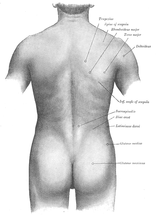
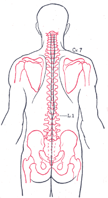
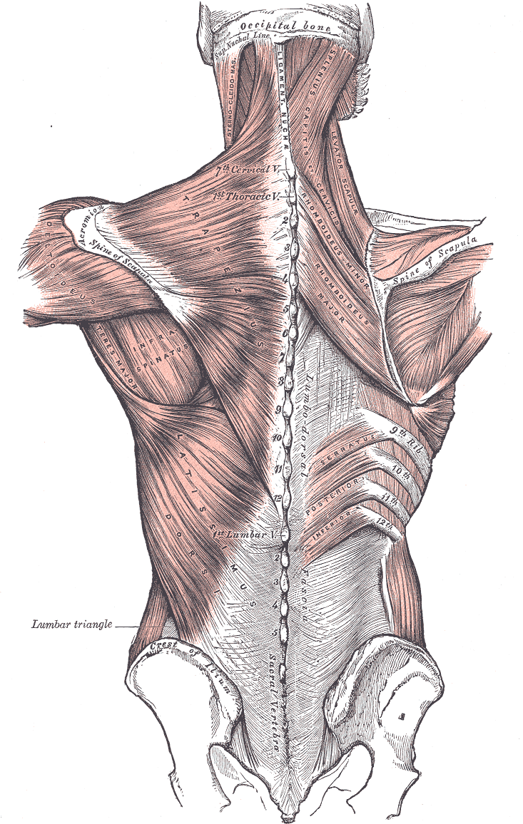
The back muscles are arranged in layers. The superficial layer controls the scapula and shoulder; the deep layer (erector spinae) extends and rotates the vertebral column.
The trapezius is a large, diamond-shaped muscle that covers the upper back, extending from the skull and cervical vertebrae to the scapula and clavicle. It retracts, elevates, and depresses the scapula. The latissimus dorsi is the broadest muscle of the back, extending the shoulder, adducting it, and internally rotating it (as in a pull-up or swimming stroke). The rhomboids (major and minor) retract the scapula, pulling it toward the midline.
The erector spinae is a deep mass of muscle running from the sacrum to the skull, divided into three columns: iliocostalis (lateral), longissimus (intermediate), and spinalis (medial). It extends the vertebral column and resists forward flexion. Deep to the erector spinae are the transversospinalis muscles (semispinalis, multifidus, rotatores), which provide fine-tuning of vertebral position and proprioception.
Chest and Abdomen
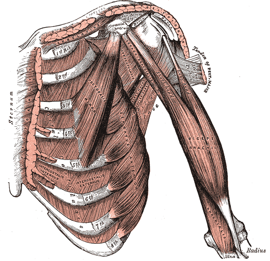
The pectoralis major is the large, fan-shaped muscle of the anterior chest that adducts, flexes, and internally rotates the shoulder. Deep to it lies the pectoralis minor, which protracts and depresses the scapula. The serratus anterior wraps around the lateral chest wall and protracts the scapula — it is known as the “boxer’s muscle” because it powers a forward punch.
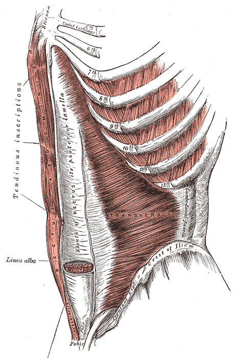
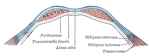
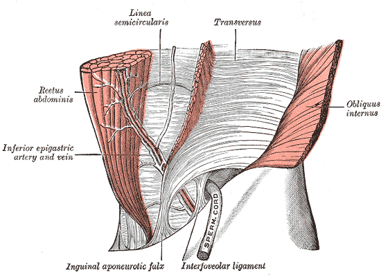
The anterior abdominal wall consists of four muscles arranged in layers. The rectus abdominis runs vertically from the pubic symphysis to the xiphoid process and costal cartilages; its tendinous intersections produce the “six-pack” appearance. The external oblique (outermost) fibers run downward and medially; the internal oblique fibers run upward and medially; and the transversus abdominis (deepest) fibers run horizontally. Together, they compress the abdomen, flex and rotate the trunk, and protect the abdominal viscera.
Shoulder
The deltoid caps the shoulder joint and is the primary abductor of the arm. Its anterior fibers flex and internally rotate, its middle fibers abduct, and its posterior fibers extend and externally rotate.
The rotator cuff consists of four muscles whose tendons fuse with and reinforce the shoulder joint capsule. The supraspinatus initiates abduction, the infraspinatus and teres minor externally rotate, and the subscapularis internally rotates. The rotator cuff provides dynamic stability to the highly mobile glenohumeral joint.
Arm
The biceps brachii has two heads (long and short) arising from the scapula and inserting on the radial tuberosity. It flexes the elbow and supinates the forearm. The brachialis lies deep to the biceps and is the primary flexor of the elbow. The triceps brachii has three heads and is the sole extensor of the elbow.
Forearm and Hand
The forearm contains two compartments separated by the interosseous membrane and fascia. The anterior (flexor) compartment contains the wrist and finger flexors, innervated primarily by the median nerve. The posterior (extensor) compartment contains the wrist and finger extensors, innervated by the radial nerve.
The intrinsic muscles of the hand are divided into three groups. The thenar muscles (abductor pollicis brevis, flexor pollicis brevis, opponens pollicis) control thumb movement and are innervated by the median nerve. The hypothenar muscles control the little finger and are innervated by the ulnar nerve. The lumbricals and interossei control the fine movements of finger flexion and extension.
Pelvis and Hip
The gluteus maximus is the largest muscle in the body and the primary extensor of the hip. It is essential for rising from a seated position, climbing stairs, and running. The gluteus medius and gluteus minimus abduct and internally rotate the hip and are critical for pelvic stability during single-leg stance (the Trendelenburg test).
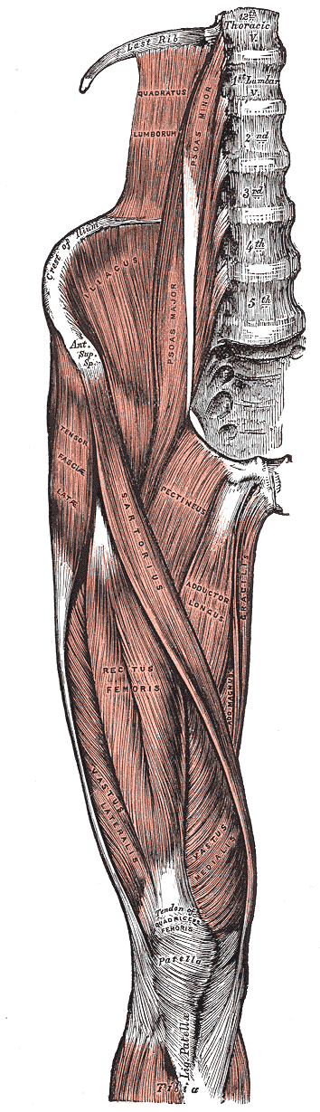
The iliopsoas (psoas major + iliacus) is the primary flexor of the hip. The adductors (adductor longus, brevis, and magnus; gracilis; pectineus) adduct the thigh and are innervated by the obturator nerve.
Thigh
The thigh is divided into three compartments by fascial septa. The anterior compartment contains the quadriceps femoris and the sartorius. The medial compartment contains the adductors. The posterior compartment contains the hamstrings.
The quadriceps femoris is the largest muscle group in the body by mass and the primary extensor of the knee. It has four heads: the rectus femoris (which also flexes the hip) and the three vasti (vastus medialis, intermedius, and lateralis). All four converge onto the patella via the quadriceps tendon, and from the patella to the tibial tuberosity via the patellar tendon.
The hamstrings consist of three muscles on the posterior thigh: the semitendinosus, semimembranosus (medial), and biceps femoris (lateral). All three extend the hip and flex the knee. The hamstrings are two-joint muscles and are prone to strain because they are stretched across both the hip and knee during activities like sprinting.
Leg and Foot
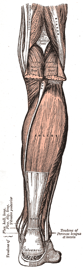
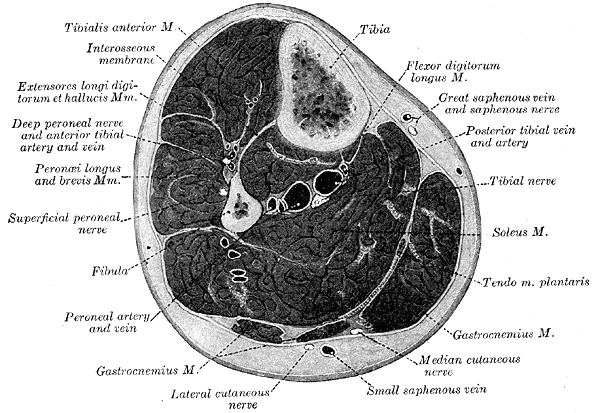
The leg is divided into four compartments. The anterior compartment contains the dorsiflexors of the ankle (tibialis anterior, extensor hallucis longus, extensor digitorum longus) and is innervated by the deep peroneal nerve. The lateral compartment contains the evertors (peroneus longus and brevis), innervated by the superficial peroneal nerve. The superficial posterior compartment contains the plantarflexors (gastrocnemius, soleus, plantaris), innervated by the tibial nerve. The deep posterior compartment contains the tibialis posterior, flexor hallucis longus, and flexor digitorum longus, also innervated by the tibial nerve.
The intrinsic muscles of the foot are arranged in four layers and control the fine movements of the toes and the maintenance of the foot arches.
Neuromuscular Junction
Every skeletal muscle fiber is innervated by a single alpha motor neuron whose cell body lies in the anterior horn of the spinal cord. The point of communication between the nerve terminal and the muscle fiber is the neuromuscular junction (NMJ) .
The motor neuron branches into many terminal boutons, each of which sits in a shallow depression on the muscle fiber surface called the motor end plate. The nerve terminal is separated from the muscle membrane by a narrow gap of approximately 50 nanometers — the synaptic cleft.
When an action potential reaches the nerve terminal, voltage-gated calcium channels open, calcium enters the terminal, and synaptic vesicles fuse with the presynaptic membrane, releasing acetylcholine (ACh) into the synaptic cleft. ACh diffuses across the cleft and binds to nicotinic receptors on the motor end plate — ion channels that open in response to ACh, allowing sodium ions to enter the muscle fiber. The resulting depolarization (end-plate potential) triggers an action potential that propagates along the muscle fiber membrane, initiating the chain of events that leads to contraction.
A motor unit consists of one alpha motor neuron and all the muscle fibers it innervates. The number of fibers per motor unit varies with the precision of movement required. In the extraocular muscles, a single motor neuron innervates only 3–5 fibers, allowing exquisitely fine control of eye position. In the quadriceps, a single motor neuron may innervate 1000–2000 fibers, producing powerful contractions with relatively coarse control.
| Muscle Type | Fibers per Motor Unit | Function |
|---|---|---|
| Extraocular | 3–5 | Very fine control (eye movements) |
| Hand intrinsics | 10–100 | Fine motor control |
| Most limb muscles | 300–1000 | Functional movement |
| Quadriceps, gluteus | 1000–2000 | Force production |
Cardiac Muscle
Cardiac muscle is found exclusively in the heart. It is striated like skeletal muscle but differs in several fundamental ways.
Cardiac muscle cells (cardiomyocytes) are short, branched, and connected end-to-end by intercalated discs. These specialized junctions contain gap junctions (which allow ions to pass directly between cells, enabling rapid electrical spread of depolarization) and desmosomes (which provide mechanical adhesion, preventing the cells from pulling apart under the constant mechanical stress of contraction).
Cardiac muscle is autorhythmic — it generates its own action potentials spontaneously, driven by the sinoatrial (SA) node, the heart’s natural pacemaker. The long refractory period of cardiac muscle prevents tetanus, which would be fatal: the heart must relax between contractions to refill with blood.
Unlike skeletal muscle, cardiac muscle has no motor units. Instead, all cardiomyocytes contract as a single functional unit (a syncytium) in response to each electrical impulse. This all-or-none response ensures coordinated ejection of blood from the chambers.
Smooth Muscle
Smooth muscle lines the walls of all hollow organs and tubular structures in the body: blood vessels, airways, the gastrointestinal tract, the ureters, the bladder, the uterus, and the iris of the eye. Its name comes from its lack of striations — the actin and myosin filaments are organized more loosely than in striated muscle and are not arranged into sarcomeres.
Smooth muscle exists in two functional types. Multi-unit smooth muscle consists of individual units that contract independently, each innervated by its own nerve terminal. This type is found in the iris, the ciliary body of the eye, and the walls of large airways. Single-unit (visceral) smooth muscle is far more common. Its cells are electrically coupled by gap junctions, so they contract as a coordinated sheet. Rhythmic, spontaneous contractions are common in single-unit smooth muscle, driven by pacemaker cells. It is found in the walls of the gastrointestinal tract, uterus, and small blood vessels.
Smooth muscle contractions are slower and more sustained than skeletal muscle contractions. An important adaptation is the latch state — smooth muscle can maintain tension for extended periods with very low ATP consumption, which is essential for organs like the bladder (which must remain contracted for hours) and blood vessels (which maintain constant tone throughout life).