The human brain is the most complex structure in the known universe. Weighing approximately 1.4 kg and containing an estimated 86 billion neurons, it is the seat of consciousness, thought, emotion, memory, and the coordinator of every voluntary and involuntary function in the body.
Major Divisions of the Brain
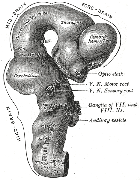
The brain is divided into five major regions during embryonic development, each persisting in the adult brain with distinct structures and functions:
- Telencephalon (cerebrum) — the largest region, responsible for higher cognitive functions
- Diencephalon — thalamus, hypothalamus, epithalamus, subthalamus
- Mesencephalon (midbrain) — part of the brainstem
- Metencephalon — pons and cerebellum
- Myelencephalon (medulla oblongata) — the lowest part of the brainstem
The Cerebrum
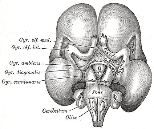
The cerebrum occupies the anterior and middle cranial fossae and constitutes about 85% of the brain’s weight. It is divided into two cerebral hemispheres connected by the corpus callosum, a massive commissural tract containing over 200 million axons.
Lobes of the Cerebrum
Each cerebral hemisphere is divided into four anatomically defined lobes, named for the overlying skull bones, plus a fifth region:
Frontal lobe. The largest lobe, lying anterior to the central sulcus. It governs executive functions (planning, decision-making, inhibition), voluntary motor control (primary motor cortex in the precentral gyrus), and speech production (Broca’s area in the dominant hemisphere).
Parietal lobe. Posterior to the central sulcus, extending to the parieto-occipital sulcus. It processes somatosensory information (touch, pain, temperature, proprioception) in the primary somatosensory cortex (postcentral gyrus) and integrates sensory input for spatial awareness and attention.
Temporal lobe. Inferior to the lateral sulcus (Sylvian fissure). It houses the primary auditory cortex, Wernicke’s area (speech comprehension), the hippocampus (memory formation), and the amygdala (emotion).
Occipital lobe. The most posterior lobe, primarily devoted to vision. The primary visual cortex (calcarine sulcus) receives input from the retinas and begins the process of visual perception.
Insula. A deep-lying lobe hidden within the lateral sulcus, involved in interoception (awareness of bodily states), pain perception, and emotional awareness.
Gyri and Sulci
The surface of the cerebrum is folded into ridges (gyri) separated by grooves (sulci or fissures). This folding dramatically increases the surface area of the cerebral cortex — approximately 2,500 cm² — allowing for greater cognitive capacity within the confines of the skull.
Key landmarks include:
- Central sulcus — separates frontal and parietal lobes; the boundary between motor and somatosensory cortices
- Lateral sulcus (Sylvian fissure) — separates temporal lobe from frontal and parietal lobes
- Parieto-occipital sulcus — marks the boundary between parietal and occipital lobes
- Calcarine sulcus — on the medial surface of the occipital lobe; contains the primary visual cortex
- Cingulate sulcus — lies above the cingulate gyrus, part of the limbic system
The Cerebellum
The cerebellum (little brain) occupies the posterior cranial fossa, inferior to the occipital lobes. Despite comprising only 10% of the brain’s volume, it contains more than half of all neurons. Its functions include:
- Coordination of movement — fine-tuning the timing, force, and trajectory of voluntary movements
- Balance and equilibrium — integrating vestibular, visual, and proprioceptive information
- Motor learning — acquiring and refining motor skills (riding a bicycle, playing an instrument)
- Cognitive contributions — increasingly recognized involvement in language, attention, and emotional regulation
The cerebellum has a highly regular, three-layered cortex and deep cerebellar nuclei (dentate, emboliform, globose, and fastigial). It connects to the brainstem via three pairs of peduncles: superior (midbrain), middle (pons), and inferior (medulla).
The Brainstem
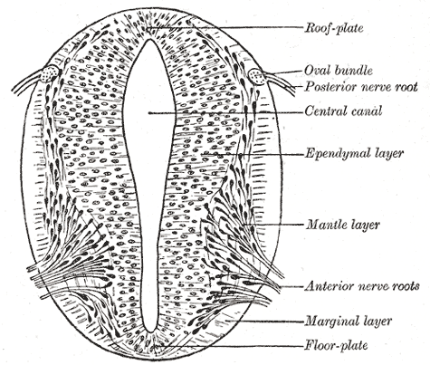
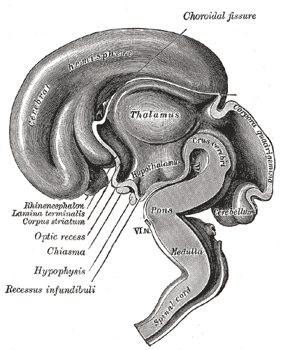
The brainstem connects the cerebrum and cerebellum to the spinal cord. It contains ascending and descending tracts, cranial nerve nuclei, and vital autonomic centers.
Midbrain (mesencephalon). The most rostral portion of the brainstem. It contains the cerebral peduncles (motor tracts), the tectum (superior and inferior colliculi for visual and auditory reflexes), and the substantia nigra (dopaminergic neurons critical for movement). The cerebral aqueduct runs through the midbrain, connecting the third and fourth ventricles.
Pons. The middle portion of the brainstem, appearing as a prominent bulge on the anterior surface. It contains nuclei that relay information between the cerebrum and cerebellum, the pontine respiratory centers, and the nuclei of cranial nerves V through VIII.
Medulla oblongata. The most caudal portion, continuous with the spinal cord at the foramen magnum. It contains the pyramids (corticospinal tracts), the decussation of the pyramids (where most motor fibers cross), and vital autonomic nuclei: the cardiovascular center (heart rate, blood pressure), the respiratory center (rhythm generation), and the nuclei of cranial nerves IX through XII.
Deep Structures of the Cerebrum
Beneath the cerebral cortex lie several crucial structures:
Basal ganglia. A group of interconnected subcortical nuclei (caudate, putamen, globus pallidus, substantia nigra, subthalamic nucleus) that regulate voluntary movement. They function as a gating system, selecting desired movements and suppressing unwanted ones. Damage to the basal ganglia produces movement disorders: Parkinson’s disease (hypokinesia), Huntington’s disease (hyperkinesia), and hemiballismus.
Thalamus. A paired, egg-shaped structure that serves as the brain’s sensory relay station. All sensory information except olfaction synapses in the thalamus before reaching the cortex. It also plays roles in motor control, arousal, and consciousness.
Hypothalamus. A small but critically important region below the thalamus. It regulates homeostasis: body temperature, hunger, thirst, sleep-wake cycles, and endocrine function via the pituitary gland. It is the major output pathway of the limbic system.
Limbic system. A functional network of structures including the hippocampus, amygdala, cingulate gyrus, and hypothalamus. It governs emotion, memory, motivation, and social behavior. The hippocampus is essential for forming new declarative memories; the amygdala processes fear and emotional salience.
Ventricles and Cerebrospinal Fluid
The brain contains four interconnected cavities (ventricles) filled with cerebrospinal fluid (CSF):
- Lateral ventricles (first and second) — large C-shaped cavities within each cerebral hemisphere
- Third ventricle — a narrow midline cavity within the diencephalon
- Fourth ventricle — between the brainstem and cerebellum, continuous with the central canal of the spinal cord
CSF is produced by the choroid plexus within the ventricles. It circulates from the lateral ventricles through the interventricular foramina (of Monro) into the third ventricle, then through the cerebral aqueduct (of Sylvius) into the fourth ventricle, and exits through the median and lateral apertures into the subarachnoid space. CSF cushions the brain, provides buoyancy (effectively reducing the brain’s weight from 1.4 kg to about 50 g), removes metabolic waste, and maintains ionic balance.
Meninges
The brain and spinal cord are wrapped in three protective membranes:
Dura mater. The tough, outermost layer. It consists of two layers (periosteal and meningeal) that are fused except where they separate to form the dural venous sinuses. Dural folds (falx cerebri, tentorium cerebelli, falx cerebelli) partition the cranial cavity.
Arachnoid mater. The delicate, web-like middle layer. It is separated from the pia mater by the subarachnoid space, which contains CSF and the major cerebral arteries.
Pia mater. The thin, vascular layer that adheres tightly to the surface of the brain and spinal cord, following every contour.
Blood Supply of the Brain
The brain receives about 15% of the cardiac output and consumes 20% of the body’s oxygen. It is supplied by two paired arterial systems:
Internal carotid arteries. These arise from the common carotid arteries and supply the anterior and middle portions of the cerebral hemispheres through the anterior and middle cerebral arteries.
Vertebral arteries. These arise from the subclavian arteries, ascend through the transverse foramina of the cervical vertebrae, and unite to form the basilar artery. The basilar artery supplies the brainstem and cerebellum and then divides into the posterior cerebral arteries.
The circle of Willis is a crucial anastomotic ring at the base of the brain, formed by the anterior and posterior communicating arteries connecting the carotid and vertebrobasilar systems. This arrangement provides collateral blood flow; if one artery is occluded, blood can reach the affected territory through the communicating vessels.
Summary
The brain is organized hierarchically, from the highly folded cerebral cortex that governs consciousness and cognition, through the deep structures that regulate movement and emotion, to the brainstem that controls vital functions. The cerebellum fine-tunes movement, the ventricles produce and circulate CSF, and the meninges and blood-brain barrier provide protection. The circle of Willis ensures a robust blood supply, while the blood-brain barrier maintains the precise chemical environment neurons require. This anatomical foundation is essential for understanding the clinical neurology that follows.